
Why Dentists Take X-rays and CT Scans: Types, Radiation and Frequency
Published: 2025-05-23 · Updated: 2026-11-22 · Medically reviewed by Katsuaki Takeuchi, DDS (Director; Board-certified oral surgeon, Japanese Society of Oral and Maxillofacial Surgeons)
What you will learn
- Why dental X-rays are taken and what they show
- The difference between intraoral, panoramic and dental CT
- How to think about radiation dose, how often X-rays are retaken, and cautions in pregnancy
Dental clinics often take X-rays during examinations. You may wonder why X-rays are needed every time, or whether the radiation is safe.
Many diseases of the mouth cannot be found just by looking. X-rays let us check the parts we cannot see, so we can make an accurate diagnosis and treatment plan. In this article, we explain the role and types of dental X-rays and how to think about radiation.
Why dental X-rays are taken
X-rays (radiographs) let us check the following.
- Decay hidden between teeth or under fillings and crowns, and how deep it is
- The shape and number of tooth roots, and whether the nerve remains
- Pus or inflammation at the root tip (periapical lesions)
- Tartar hidden under the gums, and how much bone has been lost to gum disease
- The position of wisdom teeth and teeth buried in the bone
- Abnormalities such as cysts in the jawbone
It is not unusual for what seemed to be a small cavity to turn out, once treatment begins, to have spread deep inside. Decay under a crown cannot be seen and may be found late. Checking with X-rays helps prevent such oversights and find problems while they are small. X-rays are also used to check root length and follow up after root canal treatment, and to assess the balance of teeth and jaws in orthodontics.
Types of dental X-rays
Intraoral (small) X-rays
A small film or sensor is placed in the mouth to take a detailed image of a few teeth. This is suited to seeing how deep decay is, the condition of the root tip, and fine changes in the bone.
Panoramic (whole-mouth) X-rays
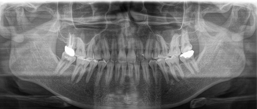
The machine rotates around your face to capture all the upper and lower teeth and the jawbones in one image. It lets us check at once the overall state of decay and gum disease, the position of wisdom teeth, and the condition of the jaw joints and bone.
Dental CT (3D examination)
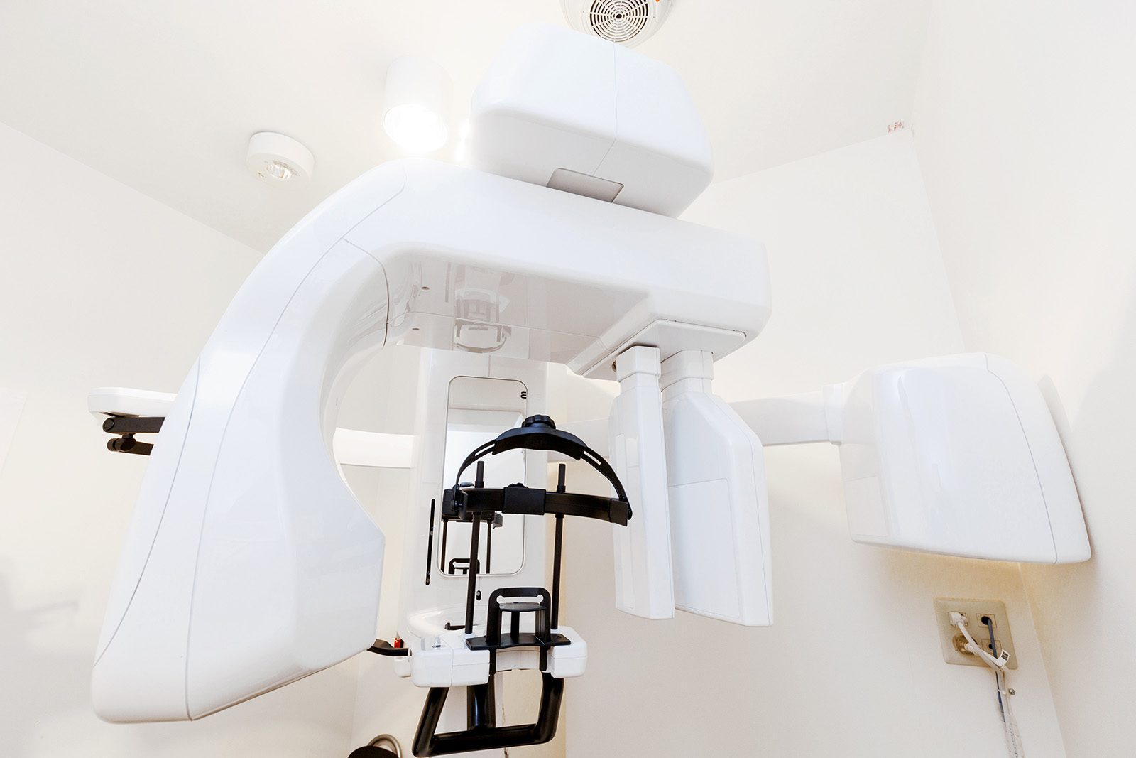
Ordinary X-rays are flat images, but dental CT shows the teeth and jawbones in three dimensions. It is useful for checking bone thickness and height and the position of nerves and blood vessels before implant treatment, the direction and shape of wisdom teeth and buried teeth and their distance from nerves, and the root shape and extent of disease in teeth needing root canal treatment. Our clinic has dental CT, so you can have these examinations in our clinic without going to a university hospital.
Is radiation safe? How to think about dose
X-rays involve a small amount of radiation exposure. However, dental X-rays cover a narrow area and the dose is very small. In daily life, we all receive natural radiation from the ground, space, food and so on, said to be about 2 mSv (millisieverts) a year in Japan. In comparison, a single intraoral or panoramic dental X-ray is said to be about one hundredth of that or less. Dental CT involves more than ordinary X-rays, but it is said to use a smaller dose than medical CT.
During imaging, you wear a protective apron. At our clinic, we take images only when needed and only of the area needed.
If you are or may be pregnant
Dental X-rays are taken far from the abdomen with a very small dose, so they are thought to have almost no effect on an unborn baby. Even so, if you are or may be pregnant, please tell us before imaging. We will consider the need and discuss whether and when to take images.
How often are X-rays taken?
At regular check-ups, X-rays are retaken at set intervals to check for new decay and the progress of gum disease. At our clinic, we update whole-mouth X-rays about once every 2 years as a guide. The interval may change depending on the condition of your teeth, your risk of decay, and whether any teeth are under treatment. Check-ups are recommended about once every 3 months, and together with X-rays, we will keep track of your mouth's condition.
Summary
Dental X-rays are an important examination for finding decay and root disease that cannot be seen, and bone changes from gum disease. We use intraoral, panoramic and dental CT according to the purpose, and keep the radiation dose to what is needed. If you have any concerns about radiation or imaging, please feel free to ask.
ESCA Dental & Orthodontic Clinic is in the ESCA underground shopping mall, a 1-minute walk from the Shinkansen exit of JR Nagoya Station. Appointments by phone (+81-52-452-1255), online booking (Japanese) or LINE.
Main risks and side effects
- X-ray and CT imaging involves a small amount of radiation exposure
- Metal fillings and crowns may distort the image and make it harder to see
Frequently asked questions
Q. Does having many X-rays affect my body?
The radiation dose of dental X-rays is very small, said to be slight even compared with the natural radiation received in a year. We take images only when needed and only of the area needed.
Q. Can I have X-rays during pregnancy?
Because the area imaged is far from the abdomen, there is thought to be almost no effect, but please tell us before imaging. We will consider the need and discuss the timing.
Q. When is CT used?
It is used to check the bone before implant treatment, the position of wisdom teeth and buried teeth, and the root shape of teeth that need root canal treatment.
Related pages
Medical review
This article was reviewed by Katsuaki Takeuchi, DDS (Director; Board-certified oral surgeon, Japanese Society of Oral and Maxillofacial Surgeons), ESCA Dental & Orthodontic Clinic. It provides general information; please consult a dentist about your own condition.
Appointments and questions
Mon–Fri 10:00–13:00 / 14:30–19:30 · Sat & Sun 10:00–13:00 / 14:00–18:00 (open every other Sunday) · Closed on national holidays. Our staff mainly speak Japanese.